Sunday, November 16, 2014

Final Observations

On Tuesday, November 11, I observed my Microaquarium for the last time. Here is a summary of what I saw:

Gastrotrich - counted 5
Euclanis - counted 12
Diatoms - several
Amoebas - counted 1
Vorticella - counted 3
Epalxis - several
Green Algae (Cladophora) - counted 1
Centropyxis - counted 1
Oscillatoria - counted 1
Calothrix - counted 1

These numbers may not accurately represent population sizes. I am very limited in my ability to spot organisms. Although the Microaquarium might appear small, it is a large amount of water for such small organisms. It is likely that I did not count every instance, and did not see some organisms at all that were present. It was especially difficult with organisms that live in or near soil, as the soil would often block clear images for identification.

The new organisms identified this week are pictured below.

Image 1: Oscillatoria, a filamentous cyanobacterium, identified from Handbook of Algae, pg. 378.

Image 2: a Calothrix cyanobacteria with visible heterocysts, identified from Handbook of Algae, pg. 427.

Image 3: Cladophora green algae covered in diatoms, as identified in Handbook of Algae, pg. 170. This algae is easily identified because it is one of only a few green algae that branch.


I learned a lot through these Microaquarium observations. While many people might have experienced declines in populations over time, my Microaquarium had plenty to observe every week. I would like to thank the University of Tennesse, Knoxville for the equipment and Dr. Ken McFarland for his invaluable assistance in identifying organisms every week.

Amber O'Malley

Sunday, November 9, 2014

Week 3 of Observation

This is the week after food was added to the Microaquarium. There were several population changes to note this week.

There were many more rotifers, particularly Euchlanis. I also noticed a good increase in population of Tachysoma. I noticed at least 3 Vorticella, the largest population yet. I saw another gastrotrich that looked like the one from week 1 observation. The gastrotrich still evades identification. I also noticed 3 or 4 amoebas, the largest number again. Like last week, there were incredible amounts of diatoms. One type was organized in a long filamentous shape. 

Image 1: a diatom, Pinnularia nobilis, as identified from Turtox Class-Room Chart: Diatoms, Figure B. This was the only structure like this that I saw. I could not find any literature as to why the diatoms were like this, if they did this on their own, if they formed their own sheath or inhabited an empty shell left behind by some organism. 

There were a couple of new organisms I identified this week.
Image 2: Difflugia identified from Free-Living Freshwater Protozoa: A Colour Guide, p. 95, Figure 186. I only saw one of these.

Image 3: Asterionella as identified from Pond III: Photosynthetic Microlife. I saw several of these diatoms. Not all had 5 cells; some had less.

Until next week.

Friday, October 31, 2014

Week 2: Feeding the Fishes

Well, technically there are no fish in my Microaquarium. But this week did bring the addition of food! On Friday, October 24, one pellet of "Atison's Betta Food" was added. See below for information about this food.

"Atison's Betta Food" made by Ocean Nutrition, Aqua Pet Americas, 3528 West 500 South, Salt Lake City, UT 84104. Ingredients: Fish meal, wheat flower, soy meal, krill meal, minerals, vitamins and preservatives. Analysis: Crude Protein 36%; Crude fat 4.5%; Crude Fiber 3.5%; Moisture 8% and Ash 15% (McFarland 2014).
Information can also be found at the Ocean Nutrition website.

I made observations of my aquarium on Tuesday, October 28th. 


There were air bubbles around the plants I added, a good sign that the plants are doing well and producing oxygen for the Microaquarium. In my observations, I saw a large number of pennate diatoms - even more than last week! I saw another vorticella, multiple amoebas, and more Litonotus cygnus. This week, I made a couple of videos to show the motion of some organisms. 


This video is of the movement of an amoeba.


This is a new organism I identified this week. This belongs to the genus Tachysoma, as identified in Free-Living Freshwater Protozoa: A Colour Guide on pg. 125, Figure 265. The video shows the movement of cilia at the end of the organism, shown by a faint flickering of the image to the right of the organism.


I spotted another rotifer this week. This is a member of the genus Euchlanis as identified in Fresh-Water Invertebrates of the United States: Protozoa to Mollusca on pages 182 and 183. I saw a couple of these.

There was a mystery organism in my aquarium this week that defied identification. It could be a juvenile form of an organism that is not as easy to identify. I will be w
atching next week to see if this organism can be identified and how it changes. It will also be interesting to see what the addition of food does, if anything, to the populations of microorganisms.


Sunday, October 26, 2014

Week 1 Observation

A week ago, the Microaquarium was set up (This is how I did it.). Once again, I examined living organisms in my Microaquarium with a microscope, but this time I was able to take pictures through the microscope. For many organisms, the pictures did not give enough detail to determine which species the organism was. I will give as much information as I can gather from my images. Here are some of the organisms I saw:

Image 1: an amoeba from the genus Mayorella, identified from Free-Living Freshwater Protozoa: A Colour Guide, p. 98, Figure 192. I saw a couple of these this week.

Image 2: a pennate diatom from the genus Navicula, identified from Free-Living Freshwater Protozoa: A Colour Guide, p, 25, Figure 5. Diatoms were plentiful in my aquarium. I also recognize these from the set-up week.

Image 3: a gastrotrich, identified from Fresh-Water Invertebrates of the United States: Protozoa to Mollusca, p. 158, Figure 1. This was the only gastrotrich I saw.

Image 4: a protozoa of the genus Urocentrum, identified from Free-Living Freshwater Protozoa: A Colour Guide, p, 167, Figure 388. There were many protozoa-like organisms in my aquarium.

Image 5: another pennate diatom from the genus Cocconeis, identified from Guide to Microlife, p. 112. 

Image 6: Litonotus cygnus, identified from Free-Living Freshwater Protozoa: A Colour Guide, p. 133, Figure 287. I saw a couple of these in this week's observation.

Image 7: a protist from the genus Actinophrys, identified from Guide to Microlife, p. 83. I only saw one or two of these.

Image 8: a protist from the genus Vorticella, identified from Guide to Microlife, p. 127. I recognize this from the set-up week. I did not see very many.

Image 9: a tiny animal of the rotifers of genus Collotheca, identified from Fresh-Water Invertebrates of the United States: Protozoa to Mollusca, p. 219, Figure 43b. This was the only one I saw.

While I only saw one of several organisms, it is possible that there were more that I did not see. I tried to capture images of moving organisms, but some moved too quickly and were too small for me to get pictures of. I saw some blue-green algae. Also, many organisms were along the soil line or beside plants, so this is where I spent the majority of my time looking. It took me almost an hour to get these pictures, and several others. You can find the information on the sources cited by visiting the bibliography page of my blog.

Hope you enjoyed these images. Unitl next week,
Amber O'Malley

Sunday, October 19, 2014

Bibliography

Botany 111 Fall 2014 [Internet]. Ken McFarland [cited 2014 October 19]. Available from: http://botany1112014.blogspot.com/

Patterson, D.J. 2013. Free-Living Freshwater Protozoa: A Colour Guide. 5th impression. London (UK): Manson Publishing Ltd.

Peachey, Gordon. 1998. Pond III: Photosynthetic Microlife [poster]. Canada: BioCam Communications Inc.

Pennak, Robert W. 1989. Fresh-water Invertebrates of the United States: Protozoa to Mollusca. 3rd ed. New York (NY): Wiley-Interscience Publication, John Wiley & Sons, Inc.

Rainis, Kenneth G; Russell, Bruce J. 1996. Guide to Microlife. USA: Franklin Watts.

Turtox Class-Room Chart: Diatoms [poster]. Chicago (United States of America): CCM: General Biological, Inc.

Homemaking

On Tuesday, October 14th, I set up a Microaquarium. The tank of the aquarium was composed of 2 pieces of plexiglass with 3 of the edges sealed with silicone caulk. The tank has a base and a lid, each a single piece of plexiglass with a groove cut out to fit around the tank. 

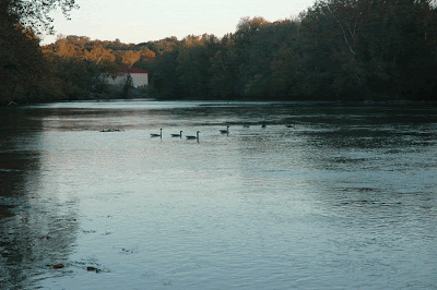
The water sample I chose is from the Holston River.


4. Holston River along John Sevier Hwy under I 40 Bridge 
Water source: Holston River along John Sevier Hwy under I 40 bridge. Partial shade exposure. Holston River water shed: N36 00.527 W83 49.549 823 ft. 10/12/2014. (McFarland 2014)

I took a few pipettes of water to fill my Microaquarium: from the bottom layer containing soil, from just above the soil, from the middle of the water container, and skimmed off the top of the water. Drawing the samples in this way ensures diversity of microorganisms.

In addition to the water, we added aquatic plants Amblestegium varium and Utricularia gibba. Both plants will oxygenate the water sample, and Amblestegium varium will consume insects and insect larvae. The sample of Holston River water had a plant in it, and I put a couple of leaves of this into my Microaquarium. 

Amblestegium varium (Hedwig) Lindberg. Moss.
Collection from: Natural spring. at Carters Mill Park, Carter Mill Road, Knox Co. TN. Partial shade exposure. N36 01.168 W83 42.832. 10/12/2014

Utricularia gibba L. Flowering plant. 

carnivorous plant. Original material from south shore of Spain Lake (N 35o55 12.35" W088o20' 47.00), Camp Bella Air Rd. East of Sparta Tn. in White Co. and grown in water tanks outside of greenhouse at Hesler
Biology Building. The University of Tennessee. Knox Co. Knoxville TN.
10/12/2014
(McFarland 2014)

To attach the top of the Microaquarium, we used two small pieces of an adhesive called Blue Stik, one on each side of the lid. Initial observations sighted multiple organisms moving in the aquarium. Some could be seen on 40x magnification, others required 100x. I examined along the soil in my tank, and did not see any organisms there.

Beginning next week, there will be weekly updates on the Microaquarium including pictures of organisms.